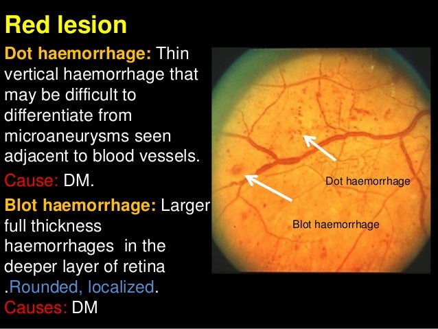
Dot And Blot Hemorrhages - Detection of N6‑methyladenosine modification residues (Review) : Dot and blot hemorrhages occur as microaneurysms rupture in the deeper layers of the retina, such as the inner nuclear and outer plexiform layers.
Dot And Blot Hemorrhages - Detection of N6‑methyladenosine modification residues (Review) : Dot and blot hemorrhages occur as microaneurysms rupture in the deeper layers of the retina, such as the inner nuclear and outer plexiform layers.. These are commonly seen in the macular area. Dot & blot vs splinter haemorrhage. Modest association with risk of clinical stroke, subclinical stroke, coronary heart disease, and death. The wisconsin epidemiologic study of diabetic retinopathy reported that background diabetic retinopathy (microaneurysms and haemorrhages) was present in nearly all subjects with type 1 diabetes of 20 years' duration, and in 80% of those with type 2 diabetes of a similar duration (2). accumulations of lipid leaks from surrounding capillaries and microaneurysms or exudates.
The capillary network in the posterior retina is found in two layers; Usually blood accumulates in the outer plexiform or inner nuclear layers, or more easily seen at peripheral retina where the nerve fiber layer is thin. You associate these hemorrhages mostly with background diabetic retinopathy. None of these underwent laser therapy for maculopathy within the study time frame (9 months from initial screening event). yellowish patches of lipid and protein within the retina.
PPT - PATHOPHYSIOLOGY OF CARBOHYDRATE METABOLISM ... from image.slideserve.com Usually blood accumulates in the outer plexiform or inner nuclear layers, or more easily seen at peripheral retina where the nerve fiber layer is thin. Fundoscopic exam cotton wool spots, hard exudates, blot and dot hemorrhages, neovascularization, flame hemorrhages, a/v nicking (the phenomenon where, on examination of the eye, a small artery (arteriole) is seen crossing a small vein (venule), which results in the compression of the vein with. Dot and blot hemorrhages occur as microaneurysms rupture in the deeper layers of the retina, such as the inner nuclear and outer plexiform layers. You associate these hemorrhages mostly with background diabetic retinopathy. Distinguishing dot blot / micro aneurysm. Dot and blot haemorrhages arise from bleeding capillaries in the middle layers of the retina. Retinal hemorrhage is basically blockage and rupturing of blood vessels of the retina. Dot blot technique can define as the process of identification of biomolecules like dna, rna or protein to detect its presence or absence in different samples taken from different cells or tissues of the individuals.
The capillary network in the posterior retina is found in two layers;
Retinal hemorrhages, often first diagnosed in the primary eye care setting, can be a presenting finding in many ocular and systemic disease states. Preretinal/vitreous or intraretinal (dot/blot) hemorrhages, microaneurysms, cotton wool spots. A dot blot (or slot blot) is a technique in molecular biology used to detect proteins. Retina is the membrane which receives light from opening of iris and only this membrane is responsible for the vision as it receives light and convert them into nerve impulses and these nerve impulses are converted into. Dot & blot vs splinter haemorrhage. The wisconsin epidemiologic study of diabetic retinopathy reported that background diabetic retinopathy (microaneurysms and haemorrhages) was present in nearly all subjects with type 1 diabetes of 20 years' duration, and in 80% of those with type 2 diabetes of a similar duration (2). Microaneurysm and blot dot hemorrhage. Blot haemorrhage cotton wool spots (cws). These are commonly seen in the macular area. Fluorescein angiography may be needed to distinguish between the two. Fundoscopic exam cotton wool spots, hard exudates, blot and dot hemorrhages, neovascularization, flame hemorrhages, a/v nicking (the phenomenon where, on examination of the eye, a small artery (arteriole) is seen crossing a small vein (venule), which results in the compression of the vein with. Dot blots have the advantage of being relatively easy to perform and result in a rapid answer as to the presence of the message. In this study, no patients with a single blot haemorrhage.
A superficial one in the nerve fibre layer and a deeper on within the inner nuclear. This page is about dot and blot hemorrhage,contains pdx ophthalmoscopy at loma linda university,vison/opthalmology at la sierra university,intraretinal microvascular abnormality (irma),lecture 7: Modest association with risk of clinical stroke, subclinical stroke, coronary heart disease, and death. Distinguishing dot blot / micro aneurysm. Ocular manifestations of systemic disease flashcards and more.
Opthalmoscopy Uploaded by Parash from image.slidesharecdn.com Distinguishing dot blot / micro aneurysm. The weakened vessels also become leaky, causing fluid to seep into the retina. Dilated fundus examination reveals unilateral disc swelling with peripapillary intraretinal hemorrhages, dilated tortuous veins, and intraretinal dot, blot, and flame hemorrhages in all quadrants, resulting in the classic blood and thunder fundus appearance (fig. Microaneurysm and blot dot hemorrhage. Abnormality of the posterior segment of the globe. Cws hard exudates (hex) and intraretinal oedema. A technique for detecting, analyzing, and identifying proteins, similar to the western blot technique but differing in that protein samples are not separated electrophoretically but are spotted through circular templates directly onto the membrane or paper substrate. Dot and blot hemorrhages occur as microaneurysms rupture in the deeper layers of the retina, such as the inner nuclear and outer plexiform layers.
Dot blot is a quick method for detecting biological samples like proteins or nucleic acids.
Dilated fundus examination reveals unilateral disc swelling with peripapillary intraretinal hemorrhages, dilated tortuous veins, and intraretinal dot, blot, and flame hemorrhages in all quadrants, resulting in the classic blood and thunder fundus appearance (fig. Retinal hemorrhages, often first diagnosed in the primary eye care setting, can be a presenting finding in many ocular and systemic disease states. accumulations of lipid leaks from surrounding capillaries and microaneurysms or exudates. Dot blot is a quick method for detecting biological samples like proteins or nucleic acids. A technique for detecting, analyzing, and identifying proteins, similar to the western blot technique but differing in that protein samples are not separated electrophoretically but are spotted through circular templates directly onto the membrane or paper substrate. None of these underwent laser therapy for maculopathy within the study time frame (9 months from initial screening event). A superficial one in the nerve fibre layer and a deeper on within the inner nuclear. If it did not resolve that. You associate these hemorrhages mostly with background diabetic retinopathy. may form a circinate pattern. Fundoscopic exam cotton wool spots, hard exudates, blot and dot hemorrhages, neovascularization, flame hemorrhages, a/v nicking (the phenomenon where, on examination of the eye, a small artery (arteriole) is seen crossing a small vein (venule), which results in the compression of the vein with. Dot & blot vs splinter haemorrhage. The wisconsin epidemiologic study of diabetic retinopathy reported that background diabetic retinopathy (microaneurysms and haemorrhages) was present in nearly all subjects with type 1 diabetes of 20 years' duration, and in 80% of those with type 2 diabetes of a similar duration (2).
It represents a simplification of the western blot method, with the exception that the proteins to be detected are not first separated by electrophoresis. These appear similar to microaneurysms if they are small; Fundoscopic exam cotton wool spots, hard exudates, blot and dot hemorrhages, neovascularization, flame hemorrhages, a/v nicking (the phenomenon where, on examination of the eye, a small artery (arteriole) is seen crossing a small vein (venule), which results in the compression of the vein with. accumulations of lipid leaks from surrounding capillaries and microaneurysms or exudates. Retinal hemorrhages, often first diagnosed in the primary eye care setting, can be a presenting finding in many ocular and systemic disease states.
Retinal microanatomy. Schisis cavities are in same layer ... from www.researchgate.net Dots are produced by the intersection after a 90° rotation between. Modest association with risk of clinical stroke, subclinical stroke, coronary heart disease, and death. This dot blot analysis experiment allows students to run their own dot blot and use it as a diagnostic tool. It represents a simplification of the western blot method, with the exception that the proteins to be detected are not first separated by electrophoresis. None of these underwent laser therapy for maculopathy within the study time frame (9 months from initial screening event). They may look like microaneurysms if small enough. may form a circinate pattern. Dot and blot haemorrhages arise from bleeding capillaries in the middle layers of the retina.
Retina is the membrane which receives light from opening of iris and only this membrane is responsible for the vision as it receives light and convert them into nerve impulses and these nerve impulses are converted into.
Microaneurysm and blot dot hemorrhage. None of these underwent laser therapy for maculopathy within the study time frame (9 months from initial screening event). Dot and blot haemorrhages arise from bleeding capillaries in the middle layers of the retina. These are commonly seen in the macular area. Retina is the membrane which receives light from opening of iris and only this membrane is responsible for the vision as it receives light and convert them into nerve impulses and these nerve impulses are converted into. Cws hard exudates (hex) and intraretinal oedema. Preretinal/vitreous or intraretinal (dot/blot) hemorrhages, microaneurysms, cotton wool spots. accumulations of lipid leaks from surrounding capillaries and microaneurysms or exudates. Modest association with risk of clinical stroke, subclinical stroke, coronary heart disease, and death. may form a circinate pattern. It represents a simplification of the western blot method, with the exception that the proteins to be detected are not first separated by electrophoresis. Commonly seen in association with diabetic or hypertensive retinopathy, peripapillary hemorrhage in patients with normal tension glaucoma that is also called splinter hemorrhage and retinal vein occlusion. Dot blot technique can define as the process of identification of biomolecules like dna, rna or protein to detect its presence or absence in different samples taken from different cells or tissues of the individuals.
You have just read the article entitled Dot And Blot Hemorrhages - Detection of N6‑methyladenosine modification residues (Review) : Dot and blot hemorrhages occur as microaneurysms rupture in the deeper layers of the retina, such as the inner nuclear and outer plexiform layers.. You can also bookmark this page with the URL : https://reynaldakun.blogspot.com/2021/05/dot-and-blot-hemorrhages-detection-of.html
Share Awesome
Belum ada Komentar untuk "Dot And Blot Hemorrhages - Detection of N6‑methyladenosine modification residues (Review) : Dot and blot hemorrhages occur as microaneurysms rupture in the deeper layers of the retina, such as the inner nuclear and outer plexiform layers."


Belum ada Komentar untuk "Dot And Blot Hemorrhages - Detection of N6‑methyladenosine modification residues (Review) : Dot and blot hemorrhages occur as microaneurysms rupture in the deeper layers of the retina, such as the inner nuclear and outer plexiform layers."
Posting Komentar